Corporate News / Blog | Stem Cells Course - Part 3

Category Archives for "Corporate News / Blog"

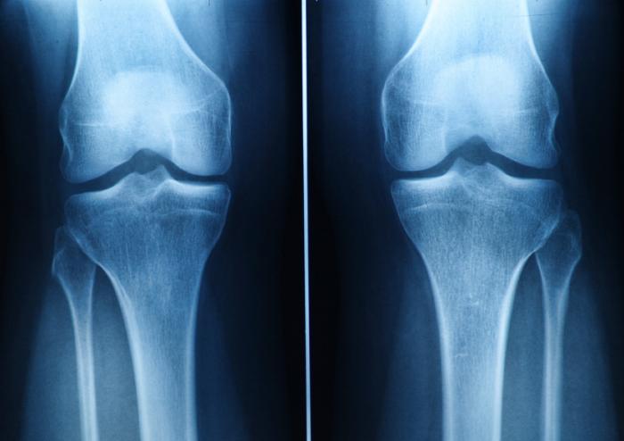
Can Stem Cells treatment Regrow the knee cartilage?

Some people believe that stem cell does all of this but studies have shown that It’s unlikely only in a few unique cases. You can observe minimal growth a year after the patient took treatment, but this doesn’t mean replacement of the cartilage.
The cartilage has a reduced regenerative capacity, and current and present pharmacological medications only offer symptomatic pain relief. Osteoarthritis patients that respond poorly to conventional therapies are ultimately treated with surgical procedures to promote cartilage repair by implantation of artificial joint structures (arthroplasty) or total joint replacement (TJR). Surgery has been the last resort for serious cartilage problems.
In the last two decades, stem cells derived from various tissues with varying differentiation and tissue regeneration potential have been used for the treatment of osteoarthritis, damage to bones and others either alone or in combination with natural or synthetic scaffolds. The stem cells derived from these tissues primarily aid cartilage repair. Although stem cells can be differentiated into chondrocytes in vitro or aid cartilage regeneration in vivo, their potential for Osteoarthritis management remains limited as cartilage regenerated by stem cells fails to fully recapitulate the structural and biomechanical properties of the native tissue. It isn’t easy for the cartilage to regrow and assume its original biomechanical and structure form.
Apparently, Due to the limited intrinsic capacity of resident chondrocytes to regrow the lost cartilage post-injury, stem cell-based therapies have been proposed as a novel therapeutic approach for cartilage repair.
Also, stem cell-based therapies using mesenchyme stem cells (MSCs) or induced pluripotent stem cells (iPSCs) have been used successfully in clinical and preclinical situations.
Part of the issues associated with Mesenchyme stem cells can be averted by using iPSCs. iPSCs are an ideal patient-specific unlimited cell source for autologous tissue regeneration. With the Promising in vitro; studies have shown that vitro results have already been demonstrated in the cartilage engineering field for iPSCs. These were generated from various cell types.
What Is Cartilage and How Does It Get Damaged?
Cartilage is a connective tissue in the human body and body of other animals. In our joints, we have a few kinds of cartilage, but most often people refer to the smooth lining of a joint called articular or hyaline cartilage. This kind of cartilage gives rise to a soft layer of cushion on the end of a bone at the joint. The cushion is essential for balance, mechanical functions and athletics. This tissue of the cartilage is very strong, yet it can compress, readjust and absorb varying degrees of energy. It is also very slippery, smooth and flexible and these features allow the joint to glide effortlessly through a broad range of physical motions of any kind.
When joint cartilage is not working correctly or damaged, this smooth-cushioning-layer can be worn away, and this becomes a problem. In the case of traumatic injuries, sometimes a sudden force causes the cartilage to break off or poorly become damaged, exposing the underlying bone of the body. In the case of osteoarthritis (also called degenerative/wear-and-tear arthritis), over time that smooth layer can wear thin and uneven. Aging can also cause the cartilage to break off and certain life factors and diseases too, e.g. autoimmune diseases.
Eventually, as that cushion of the bones wears away, joint movements can become inflexible, stiff and painful on one or both legs (bones). Joints can even become inflamed and swollen. And as all these conditions, typically causes pain and limitations in activity become problematic. The action or activities that involve these bones leads to crushing pain and discomfort, depending on the severity of the case.  Almost all activities involve the movement of bones; hence this condition is not an easy one.
There are some treatments for cartilage damage and arthritis. Although there some medicines, most of these treatments are focused either on relieving symptoms by smoothing down the damaged cartilage or concentrate on replacing the joint surface with an artificial implant. The later is for end-stage conditions, and the artificial plane is procedures such as knee replacement or hip replacement surgery.
How Can Stem Cells Help?
Stem cells are specialized cells that can multiply reform and develop into different types of tissue. In the developmental stages of a fetus, stem cells are plentiful and surplus. However, in adulthood, stem cells are restricted to specific tasks of regenerating a few types of cells, such as blood cells and liver cells in some cases of damage. There are almost no stem cells found in cartilage tissue, and therefore there is little to no capacity to heal or regrow new cartilage. For adults, the ability to regrow new cartilage is even more difficult due to age and lack of stem cells in the cartilages.
Most often, in the setting of orthopedic surgery and joint problems, stem cells are obtained from adult stem cell sources. The primary sources are bone marrow and fatty tissue. These stem cells can develop into cartilage cells, called chondrocytes.
They also exhibit some other helpful qualities by stimulating the body to reduce inflammation, stimulate cell repair, and improve blood flow. This process is caused by the secretion of cellular signals and growth factors to stimulate the body to initiate healing processes.
Once stem cells have been obtained, they need to be delivered to the area of the cartilage that damaged. One option is to inject the stem cells into the joint. There have been many studies investigating just this, and some data shows improvement in symptoms. How much of this improvement is the result of new cartilage growth versus other effects of stem cells (the healing properties listed above, including the anti-inflammatory effects) is unknown.
There is a challenge with giving stem cell injection. The problem with just injecting stem cells is that cartilage is a complex tissue that is comprised of more than only cells hence this can pose a challenge because the stem can’t regenerate all the things in the cartilage.
To regrow the cartilage, the complex tissue structure and biomechanics of cartilage must also be reconstructed to its former status. Cartilage can often /described as having a scaffold-like structure that is composed of water, cells, collagen, and proteoglycans, and infection-fighting antibodies.  Injecting just the stem cells is thought to be less effective in stimulating the formation of the entire cartilage structure hence the challenge.
Some studies are investigating the types of 3-dimensional tissue scaffolds engineered to have a cartilage-like structure. The stem can then be injected into the scaffold, in hopes of better restoring a healthy type of cartilage. Three-dimensional printing is becoming an exciting part of this type of research. If everything works out as expected, the cartilage reconstruction could be achieved to a very high percentage.
How do stem cells work?
Necessarily, stem cells are progenitor cells which are capable of regeneration and differentiation into a wide range of specialized cell types. Once injected, stem cells follow inflammatory signals from damaged tissues and have multiple ways of repairing these damaged areas. It works as though the part is developing new; like what is seen during a child’s development.
The mesenchyme stem cells (MSCs) we are using are considered to be multipotent (they can transform into different cell types but cannot form an organ) but not pluripotent. In the body, these cells Do NOT function by transforming into different cell types or tissues.
They act via anti-inflammatory activity, immune modulating capacity, and the ability to stimulate regeneration.   We go through a very high thorough screening process to find cells that we know have the best anti-inflammatory activity, the best immune modulating capacity, and the best ability to stimulate regeneration process on the tissue with damage.
ISSCA (International Society for Stem Cells Applications) www.issca.us 
This is a business located in Miami, FL, where people around the world come to take a certification in the newest Stem Cells Protocols.
Some organizations have put in efforts to help discover some solutions in stem medicine. International Society for Stem Cell Application (ISSCA ) is one of the leading associations in setting standards and promoting excellence in the field of Regenerative Medicine, researches, publications related education, certification, research and publications.
The ISSCA is a unique-multidisciplinary community of physicians, stem specialist and scientists with a mission to advance the science, technology and practice of Regenerative Medicine. Their aim is to treat disease and lessen human suffering. ISSCA generally advances the specialty of Regenerative Medicine and serves its members.
The ISSCA provides certifications and standards in the practice of Regenerative Medicine as a medical specialty.
Although the expectation on this stem cell course is yet to be achieved; however, this is a part of medicine that can offer one-end-solution to various bone and body problems.
With the recent high-tech studies, efforts and dynamics, stem cell treatment can be a breakthrough in the future as its perspectives are very promising and unique. It is also not dangerous on the long-run.

Clinical trials on NK cells give hope for many people Who are suffering from cancer.

Induced pluripotent stem cells (also known as iPS cells or iPSCs) are a type of pluripotent stem cell that can be generated directly from a somatic cell. Pluripotent stem cells hold promise in the field of regenerative medicine. Because they can propagate indefinitely, as well as give rise to every other cell type in the body (such as neurons, heart, pancreatic, and liver cells), they represent a single source of cells that could be used to replace those lost to damage or disease.

Natural killer cells are the type of cytotoxic lymphocyte critical to the innate immune system. The role NK cells play is analogous to that of cytotoxic T cells in the vertebrate adaptive immune response. NK cells provide rapid responses to virus-infected cells, acting at around three days after infection, and respond to tumor formation. 

Typically, immune cells detect the major histocompatibility complex (MHC) presented on infected cell surfaces, triggering cytokine release, causing apoptosis. NK cells are unique, however, as they can recognize stressed cells in the absence of antibodies and MHC, allowing for a much faster immune reaction.

Clinical Trial on NK cells

In a first clinical trial, a natural killer cell immunotherapy derived from induced pluripotent stem cells is being tested for safety in 64 patients with a variety of solid tumors. The first subjects used for the study received the cells in February at the University of California, San Diego (UCSD) Moores Cancer Center and MD Anderson Cancer Center. 

This study is targeting late-stage cancer patients with solid tumors, including lymphoma, colorectal cancer, and breast cancer. The FT500 NK cells do not undergo any further alterations and after their derivation from the induced pluripotent stem cells (iPSCs), offering the possibility of a quicker, ready-made treatment. 

Human embryonic stem cells induced iPSCs 

Human embryonic stem cells (hESCs) and induced pluripotent stem cells (iPSCs) provide an accessible, genetically tractable, and homogenous starting cell population to efficiently study human blood cell development. These cell populations provide platforms to develop new cell-based therapies to treat both malignant and nonmalignant hematological diseases.

The NK cells are immune cells in the same family as T and B cells and are very good at targeting cancer cells for destruction. Some Laboratory experiments have shown they do so by attacking cells that have lost their significant self-recognition signals that tell the immune system not to attack. This is the phenomenon that can happen among cancer cells but not to healthy cells. Experts are not sure how many cancer cells lose that signal. Researchers are hopeful that the clinical trial can help determine which cancer patients could benefit the most from NK cell treatment. 

iPS Clone

The ability to induce pluripotent stem cells from committed, human somatic cells provides tremendous potential for regenerative medicine. However, there is a defined neoplastic potential inherent to such reprogramming that must be understood and may offer a model for critical understanding events in the formation of the tumor. Using genome-wide assays, we identify cancer-related epigenetic abnormalities that arise early during reprogramming and persist in induced pluripotent stem cell (iPS) clones. These include hundreds of abnormal gene silencing events, patterns of aberrant responses to epigenetic-modifying drugs resembling those for cancer cells, and presence in iPS and partially reprogrammed cells of cancer-specific gene promoter DNA methylation alterations.

Progress in adoptive T-cell therapy for cancer and infectious diseases is hampered by the lack of readily available antigen-specific, human T lymphocytes. Pluripotent stem cells could provide an estimable source of T lymphocytes, but the therapeutic potential of human pluripotent stem cell-derived lymphoid cells generated to date remains uncertain.

Modification of T cells

Recently, some Approved cell therapies for Cancer also rely on modifying T cells, in those cases to produce cancer cell–binding chimeric antigen receptors (CARs), and have been effective in treating certain cancers such as leukemia.

Application of CAR T-Cell Therapy in Solid tumours

The Car T technology has wowed the field by all but obliterating some patients’ blood cancers, but solid malignancies present new challenges.

Therapies that contains such chimeric antigen receptor (CAR) T cells have been approved for some types of so-called liquid cancers of the blood and bone marrow, large B-cell lymphoma and B-cell acute lymphoblastic leukemia. But the approach has not had as much success for solid tumors.

Serious research into the therapy for brain cancer started almost 20 years ago after cancer biologist WaldemarDebinski, then at Penn State, discovered that the receptor for the immune signaling molecule interleukin 13 (IL-13) was present on glioblastomas, but not on healthy brain tissue. The receptor thus seemed like an excellent target to home in on cancer cells while sparing healthy ones. The CAR spacer domain that spans the immune cells’ membranes and its intracellular co-stimulatory areas, as well as the process used to expand cells outside the body, to boost the T cells’ activity.

CAR T- A Safer Cell Therapy

While managing CAR T-cell therapy toxicity could help keep already-designed treatments on their march to the clinic, many immunotherapy companies are also working to develop a new generation of inherently safer therapies, yet just as efficient. A crucial part of achieving this goal will be improving CAR specificity for target cells. With current treatments, the destruction of normal cells is often an unavoidable side effect when healthy tissue carries the same antigens as tumors; noncancerous B cells, for example, are usually casualties in CD19-targeted therapies.

CAR T delivery is a non-easy factor in the treatment of solid tumors and other unknown forms of tumors. With the non-solid cancers, cells are administered by a blood infusion, and once in circulation, the CAR T can seek out and destroy the rogue cells. For solid tumors, it’s not so simple.

The main drawback of taking cells from a patient and developing them into a cellular immunotherapy product is that the process can take weeks. 

Patel tells The Scientist “But for the majority of patients who may not be a candidate or may not have time to wait for such an approach, the idea that there’s off-the-shelf immunotherapy that could potentially as a living drug act against their cancer, I think is a fascinating concept,”  

Stem cell treatment could offer one-end-solution to Diabetes

Insulin-producing cells grown in the lab could provide a possible cure for the age-long disease (diabetes).

Type 1 diabetes is an auto¬immune disease that wipes out insulin-producing pancreatic beta cells from the body and raises blood glucose to dangerously high levels. These high levels of Blood sugar level can be even fatal. Patients are being administered insulin and given other medications to maintain blood sugar level. To those who cannot maintain their blood sugar level, they are given beta-cell transplants but to tolerate beta cell transplants; patients have to take immunosuppressive drugs as well.

A report by a research group at Harvard University tells us that they used insulin-producing cells derived from human embryonic stem cells (ESCs) and induced pluripotent stem cells to lower blood glucose levels in mice. Nowadays, many laboratories are getting rapid progress in human stem cell technology to develop those cells that are functionally equivalent to beta-cells and the other pancreatic cell types. Other groups are developing novel biomaterials to encapsulate such cells and protect them against the immune system without the need for immunosuppressant.

Major pharmaceutical companies and life sciences venture capital firms have invested more than $100 million in each of the three most prominent biotechnological industries to bring such treatments into clinical use:

  • Cambridge
  • Massachusetts–based companies Semma Therapeutics 
  • Sigilon Therapeutics, and ViaCyte of San Diego

Researchers of UC San Francisco have transformed human stem cells into mature insulin-producing cells for the first time, a breakthrough in the effort to develop a cure for type-1 (T1) Diabetes. Replacing these cells, which are lost in patients with T1 diabetes, has long been a dream of regenerative medicine, but until now scientists had not been able to find out how to produce cells in a lab dish that work as they do in healthy adults.

What is T1 diabetes?

T1 diabetes is an autoimmune disorder that destroys the insulin-producing beta cells of the pancreas, typically in childhood. Without insulin’s ability to regulate glucose levels in the blood, spikes in blood sugar can cause severe organ damage and eventually death. The condition can be managed by taking regular shots of insulin with meals. However, people with type 1 diabetes still often experience serious health consequences like kidney failure, heart disease and stroke. Patients facing life-threatening complications of their condition may be eligible for a pancreas transplant from a deceased donor, but these are rare, and they are supposed to wait a long time.

Researchers have just made a breakthrough that might one day make these technologies obsolete, by transforming human stem cells into functional insulin-producing cells (also known as beta cells) – at least in mice.

“We can now generate insulin-producing cells that look and act a lot like the pancreatic beta cells you and I have in our bodies,” explains one of the team, Matthias Hebrok from the University of California San Francisco (UCSF).

“This is a critical step towards our goal of creating cells that could be transplanted into patients with diabetes.”

Type-1 diabetes is characterized by a loss of insulin due to the immune system destroying cells in the pancreas – hence, type 1 diabetics need to introduce their insulin manually. Although this is a pretty good system, it’s not perfect.

Making insulin-producing cells from stem cells

Diabetes can be cured through an entire pancreas transplant or the transplantation of donor cells that produce insulin, but both of these options are limited because they rely on deceased donors. Scientists had already succeeded in turning stem cells into beta cells, but those cells remained stuck at an early stage in their maturity. That meant they weren’t responsive to blood glucose and weren’t able to secrete insulin in the right way.

Scientists at the University of California San Francisco made a breakthrough in the effort to cure diabetes mellitus type 1.

For the first time, researchers transformed human stem cells into mature insulin-producing cells, which could replace those lost in patients with the autoimmune. There is currently no known way to prevent type-1 (T1) diabetes, which destroys insulin production in the pancreas, limits glucose regulation, and results in high blood sugar levels. The condition can be managed with regular shots of insulin, but people with the disease often experience serious health complications like kidney failure, heart disease, and stroke.

“We can now generate insulin-producing cells that look and act a lot like the pancreatic beta cells you and I have in our bodies,” according to Matthias Hebrok, senior author of a study published last week in the journal Nature Cell Biology.

“This is a critical step toward our goal of creating cells that could be transplanted into patients with diabetes,” Hebrok, director of the UCSF Diabetes Center, said in a statement.

Islets of Langerhans are groupings of cells that contain healthy beta cells, among others. As beta cells develop, they have to separate physically from the pancreas to form these islets.

The team artificially separated the pancreatic stem cells and regrouped them into these islet clusters. When they did this, the cells matured rapidly and become responsive to blood sugar. In fact, the islet clusters developed in ways “never before seen” in a lab. After producing these mature cells, the team transplanted them into mice. Within days, the cells were producing insulin similar to the islets in the mice. While the study has been successful in mice, it still needs to go through more rigorous testing to see if it would work for humans as well. But the research is up-and-coming. “We can now generate insulin-producing cells that look and act a lot like the pancreatic beta cells you and I have in our bodies. This is a critical step towards our goal of creating cells that could be transplanted into patients with diabetes,” He said.

“We’re finally able to move forward on several different fronts that were previously closed to us,” he added. “The possibilities seem endless.”

Basic research keeps elucidating new aspects of beta cells; there seem to be several subtypes, so the gold standard for duplicating the cells is not entirely clear. Today, however, there is “a handful of groups in the world that can generate a cell that looks like a beta cell,” says Hebrok, who currently acts as scientific advisor to Semma and Sigilon, and has previously advised ViaCyte. “Certainly, companies have convinced themselves that what they have achieved is good enough to go into patients.”

The stem cell reprogramming methods that the three companies use to prompt cell differentiation create a mixture of islet cells. Beta cells sit in pancreatic islets of Langerhans alongside other types of endocrine cells. Alpha cells, for example, churn out glucagon, a hormone that stimulates the conversion of glycogen into glucose in the liver and raises blood sugar. Although the companies agree on the positive potential of islet cell mixtures, they take different approaches to developing and differentiating their cells. Semma, which was launched in 2014 to commercialize the Harvard group’s work and counts Novartis among its backers, describes its cells as fully mature, meaning that they are wholly differentiated into beta or other cells before transplantation. “Our cells are virtually indistinguishable from the ones you would isolate from donors,” says Semma chief executive officer BastianoSanna

To get around the donor problem, researchers, including the team at UCSF has been working on nudging stem cells into becoming fully-functional pancreatic beta cells for the last few years. Still, there have been some issues in getting them all the way there.

“The cells we and others were producing were getting stuck at an immature stage where they weren’t able to respond adequately to blood glucose and secrete insulin properly,” Hebrok said.

“It has been a major bottleneck for the field.” 

“We’re finally able to move forward on a number of different fronts that were previously closed to us,” Hebrok added. “The possibilities seem endless.” 

Regardless of starting cell type, the companies say they are ready to churn out their cells in large numbers. Semma, for example, can make more islet cells in a month than can be isolated from donors in a year in the United States, Sanna says, and the company’s “pristine” cells should perform better than donor islets, which are battered by the aggressive techniques required for their isolation.

As these products, some of which have already entered clinical trials, move toward commercialization, regulatory agencies such as the US Food and Drug Administration (FDA) and the  European Medicines Agency have expressed concern about the plasticity of the reprogrammed cells. All three firms subject their cells to rigorous safety testing to ensure that they don’t turn tumorigenic. Before successful trials, companies won’t know the dose of beta cells required for a functional cure, or how long such “cures” will last before needing to be boosted. There’ll be commercial challenges, too: while the companies are investing heavily to develop suitable industrial processes, all acknowledge that no organization has yet manufactured cell therapies in commercial volumes.

Nevertheless, there’s growing confidence throughout the field that these problems will be solved, and soon. “We have the islet cells now,” says Alice Tomei, a biomedical engineer at the University of Miami who directs DRI’s Islet Immuno-engineering Laboratory.

“These stem cell companies are working hard to try to get FDA clearance on the cells.”

Protecting stem cell therapies from the immune system

Whatever the type of cell being used, another major challenge is delivering cells to the patient in a package that guards against immune attack while keeping cells fully functional. Companies are pursuing two main strategies: 

  • Microencapsulation, where cells are immobilized individually or as small clusters, in tiny blobs of a biocompatible gel.
  • Macroencapsulation, in which greater numbers of cells are put into a much larger, implantable device.

ViaCyte, which recently partnered with Johnson & Johnson, launched its first clinical trial in 2014. The trial involved a micro-encapsulation approach that packaged up the company’s partially differentiated, ESC-derived cells into a flat device called the PEC-Encapsulation. About the size of a Band-Aid, the device is implanted under the skin, where the body forms blood vessels around it. “It has a semipermeable membrane that allows the free flow of oxygen, nutrients, and glucose,” says ViaCyte’s chief executive officer, Paul Laikind. “And even proteins like insulin and glucagon can move back and forth across that membrane, but cells cannot.”

The trial showed that the device was safe, well-tolerated, and protected from the adaptive immune system—and that some cells differentiated into working islet cells. But most cells didn’t engraft effectively because a “foreign body response,” a variant of wound healing, clogged the PEC-Encap’s membrane and prevented vascularization. ViaCyte stopped the trial and partnered with W. L. Gore & Associates, the maker of Gore-Tex, to engineer a new membrane. “With this new membrane,” says Laikind, “we’re not eliminating that foreign body response, but we’re overcoming it in such a way that allows vascularization to take place.” The company expects to resume the trial in the second half of this year, provided it receives the green light from the FDA.

Semma is also developing macro¬-encapsulation methods, including a very thin device that in prototype form is about the size of a silver dollar coin. The device is “deceptively simple, but it allows us to put [in] a fully curative dose of islets,” Sanna says.

Semma is also investigating microencapsulation alternatives. More info about all on 4 dental implants cost in Temecula you can find on www.temeculafacialoralsurgery.com/ site. At the same time, the company is advancing toward clinical trials using established transplantation techniques to administer donated cadaver cells to high-risk patients who find it particularly difficult to control their blood glucose levels. These cells are infused via the portal vein into the liver, and patients take immunosuppressive drugs to prevent rejection.

Sigilon is working on its microencapsulation technology. Launched in 2016 on the back of work by the labs of Robert Langer and Daniel Anderson at MIT, the company has created 1.5-millimeter gel-based spheres that can hold between 5,000 and 30,000 cells (Nat Med, 22:306–11, 2016). Each sphere is like a balloon, with the outside chemically modified to provide immune-protection, says Sigilon chief executive officer Rogerio Vivaldi. “The inside of the balloon is full of a gel that creates almost a kind of a matrix net where the cells reside.”

In 2018, shortly after partnering with Eli Lilly, Sigilon and collaborators published research showing that islet cells that were encapsulated in gel spheres and transplanted into macaques remained functional for four months. The company has not disclosed a time frame for a type 1 diabetes trial “but we’re moving pretty quickly,” says chief scientific officer David Moller.

Conclusion

To conclude, all three firms hope to extend their work to treat some of the 400 million people worldwide with type 2 diabetes, many of them eventually benefit from insulin injections. The recent endorsements from big Pharmaceutical underline the real progress in beta-cell transplants, says Aaron Kowalski, a molecular geneticist and chief executive officer at JDRF, a foundation based in New York that has funded research at ViaCyte and academic labs whose work has been tapped by Semma and Sigilon. “These companies all realize that if they don’t do it, somebody else will. It’s hard to predict exactly when, but somebody is going to make this work.”

Bats Carry Corona Virus. So Why Don’t They Get Sick?

A lot of viruses that has taken a toll on life, the ebola virus in Africa, The Nipah virus of Nipah and the most recent one corona virus that left china running helter skelter all seemed to have originated from bats. During the course of the virus epidemic in Wuhan where it was first detected, some Chinese researchers in Wuhan examined some patients affected in that area and then took samples of the virus.
They did findings on the genetic sequence of the virus with other viruses that were known. The corona virus surprisingly had a 96% match with the horseshoe bats that are dominant in the southwest of china. The research findings were then published in a study on February, 2020.
A virologist Vineet Menachery from the university of Texas Medical Branch at Galveston though not affiliated to the study said “They’re too close in terms of their pure genetics to say they’re not related, or that they didn’t have a common ancestor.”
Menachery was a reputable virologist and had done other research works. He contributed to the theory that the spread of the corona virus must have been from these bats to humans. And possibly must have had another animal that served as an intermediary for the spread.
This same thing had happened with other forms of corona viruses as noted in the case of SARS (Severe Acute Respiratory Syndrome) an outbreak that took place in 2002-2003 where civets, a mongoose family member were infected with the bat corona virus and spread as humans bought them for food.
Another case was the MERS (Middle East Respiratory Syndrome) outbreak. This one happened in 2012 and was as a result of infected camels from the virus. People who ate undercooked meat of camels and as well drank the raw milk of camels were all affected.
So why is it that there are so many diseases that are spread from bats?
Its no doubt, bats have a lot of viruses that they carry with them. And these viruses in their variety are spread and manifests its tolls on people. Scientist are not sure why this is the case as confirmed by Kevin Olival, a research vice president as EcoHealth Alliance, a non-profit organization based in the U.S. He went further to say that it may have something to do with the family of the viruses carried by the bats. So you know, there are over 130 different families of viruses that bats do carry around.
And then, most bats and humans do come in contact through several means. The millions of populations of bats are ubiquitous to all the continents apart from in antartica. Rebekah of Colorado State university who researched infectious pathogens said “There’s a lot of viruses we’re finding in bats because there’s a lot of bats out there.”
They move about in multitudes and live in colonies of large populations. Some these members live in caves and share caves and trees where there can be a contact between humans and bats. Hence, these viruses can spread from these bats to humans.
Despite their sizes, bats have relatively long lifespans and can live over 30 years.”So there’s a long time for them to be persistently infected with the virus and shed it into the environment,” Kading says. The mode of mechanisms for these viruses are through urine, saliva and feces of bats. The outbreak of Nivah that happened in Bangladesh was linked to the sap of a date palm gotten from some trees that some bats licked and had infested with their urine.
Reading through all these, it is not absurd to wonder why the bats themselves do not get affected by the viruses they carry.
The answer to that question is based on the fact that the bat is the only flying mammal in the world. Their body metabolism and process quite differ from that of normal mammals too. When bats fly, their heart rates rise to about a thousand beats per minute with a temperature rise of about 100 degrees Fahrenheit. Linfa Wang a student of bat viruses at Duke-NUS Medical School in Singapore says that when these signs manifest in other mammals, they are signals that can trigger death. But this is not the same case for other bats. This is a lifestyle for them, every day.
Their system is also capable of producing molecules that other organisms do not have. The molecules carry out repair functions and prevent cell damage. This makes their system a bit irresistible to infections and also make them recalcitrant to viruses and resilient to diseases such as diabetes, cancer and other health conditions.
This is a prove that the manifestation of viruses in mammals is not always as a result of the virus itself, but as a result of the body’s reaction to the presence of such a virus that makes us ill by triggering other chain reactions, as Wang explains.
Olival at EcoHealth explains that these bats have coevolved with these viruses and it is not totally their fault that we humans are infected and affected by these viruses. The actual problem is when the viruses move from their species to other species of mammals which is also fostered by human activity.
Naturally, it would be hard for most animals and mammals to cross paths. But Olivial says that the presence of some activities and availability of exchange platforms made available by humans can allow such interaction to occur. She gave an example using wildlife markets like the one in Wuhan, where a bat could be mixed up with a civet. Who later on come in contact with humans – eg. Butchers who do not observe proper hygiene and protection from animal blood.
“The way that we’re coming into contact with these animals, hunting, selling, and trading them is to a scale that really we haven’t seen before,” he says.
Investigative teams did some in-depth search and they discovered some traces of this virus in 22 stalls and in a garbage truck that was found at Huanan Seafood Market right there in Wuhan, a place known for booming trade for live animals. This discovery led to shutting down the market as it was tied to majority of the cases.
The intermediary animals to this viruses are still a mystery, but it is clear that some of these animals are prone to interact more with humans. This is why when they are infected, the likelihood for human infection is widened. These other infected animals can sneeze, urinate, be cooked as food or even owned as pets.
Bats are not just vectors for viruses, they play an important role in balancing the eco-system. They feed on insects and fruits and are active agents of pollination. In fact, Wang believes that since these bats have successfully coevolved with these viruses, there is every possibility that they can be the agents that can lead to the cure and provision of therapies for these viruses.

A New Medical Device, in the Management of Complex Wounds

Because of the complex nature of wound healing process, an injury on the skin can pose several challenges and are likely pose complications especially when they are acute. They can as well deteriorate from acute to chronic conditions which will require external intervention best understood by a specialist physician to get the area affected by the wound under normalcy.

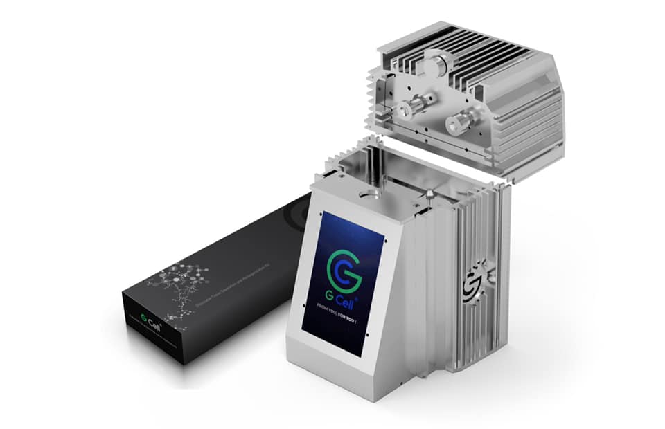
The complexity of wound healing and research remains an ocean of knowledge that is continuously researched intensely to uncover depths of wound healing techniques and interventions. Hence, this report contains an introduction and details to the use of a new medical innovation called Gcells used primarily for the management of wounds in their different etiology.

In a case where the process of wound healing seemed difficult, Gcells proved great effects an attribute to their design and working protocol. Gcells are conditioned to work with an enriched suspension of progenitor cells that can efficiently aid tissue repair process. In this case report, two subjects were used as donors and acceptors of these micro-grafts.

Introduction

The skin is an outer layer of the body, offering protection to the underlying layers. A wound breaks this layer and inhibits the various functions as well as expose or also break the underlying layer of tissues. Repair processes are inherent and part of homeostatic processes of the body to try to restore the skin back to its normalcy in structure and in function.

The basics for the skins repair mechanism is represented by a cloth and an inflammation where vessels dilate and monocytes activate leading to breakdown of necrotic tissues. This basic process can be inhibited or delayed by a number of varying factors that lead to deteriorative transformation of acute wounds to chronic forms. But if there is no alteration in the repair process, Mesenchymal cells kickstart proliferative process and begin to repair and restructure the affected tissues starting from the base. At the same time epithelial tissues begin to grow around the wound leading to a final step of the healing process. In this final stage, remodeling of the skin structure is primary and then maturation of a scar.

These processes are efficient best in certain conditions which if affected by factors such as cardiovascular ailment, diabetes, bacterial or any other genre of infection, can inhibit these processes.

Hence, it is necessary to understand in details these processes if there is going to be development or innovation for effective healing processes. Just as stated above, during the proliferative phase of wound healing, Mesenchymal cells are the key role players. Their structure includes a Mesenchymal stem cell (MSCs), multi potent in nature and offer supportive, therapeutic and trophic functions. They are also able to release viable trophic, anti-inflammatory cytokines and anti-apoptotic molecules that offer protection during the repair of wounded skin. MSCs also possess subpopulations that are stem-like nature commonly referred to as “side population” (SP) they have been found out to be enriched in over 1000-fold of progenitor cells and multipotent stem cells and as well exist in tissues and tumors. SP exists in a variety of organs and tissues, after an original discovery to be prominent in the bone marrow of a mouse. The organs with SP include the lung, liver, brain, mammary gland and in skeletal muscles.

In other discoveries, it was discovered that they probably may also be isolated in other tissues of the body. This discovery was in an in vitro and in vivo experiment when Dental Pulp Stem Cells (DPSCs) showed capability to differentiate into osteoblasts and built a woven bone by forming an Extracellular Matrix (ECM) secreted by the osteoblasts. The experiment drew results on the both the quality and quantity of the matrix formed by the DPSCs in the in vivo and in vitro experiment using Stem cells and accompanying biomaterials.

Thus proved that dental pulp holds potentialities of therapeutic strength and a rich source of progenitor/autologous cells that can be used to aid healing processes even applicable to regeneration of craniofacial bones.

This is the evidence that supports the working principle of Gcells innovation. Gcell successfully separates this side population with a size of 50 micron. At this cell population, they can form autologous micro-grafts and can either be used alone or alongside biomaterials prepared in a biocomplex ready for use when necessary.

In this case report, two subjects were used as donors and acceptors of these micro-grafts for enhanced healing of complex wounds through autologous micro-grafts using the Gcell.

Clinical case 1

The first case involves a woman at age 50 who does not have any diseases or disorders. She underwent a laparoscopic gastric bypass surgery and was doing well considering parameters of weightloss. Two years later she moved in for abdominoplasty bariatric. Later on, post complications showed preeminence of necrosis which was discovered after first medical examination were about 150 to 200cm2 at the end of the flaps. An initial necrosectomy showed an intense loss of tissue and we furthered to place the wound on VAC therapy and the patient in active participation of this therapy for one week then at home as an outpatient.

As an outpatient, there was improved and progressive wound cleansing while granulated tissues around the base area were cleared.

The VAC therapy after 2 months still left the margins of the wound deteriorated and surrounding areas not in axis with skin surface.

The Gcell protocol kickstarted after consent from the outpatient. We started by collecting a 3 cm2 skin sample from the patient for the purpose of obtaining the cell suspension needed to be injected to the granulation tissue (figure2).

We followed up with conventional wound treatment as in cleaning and replacement with sterile gauze dabbed with Vaseline. The wound area began to improve in both healing progress and general appearance. In two months, the undermined area disappeared as well as leveled to the axis of the skin surface. 2 months later, the wound reduced to a very little scar that is mild and smoothed compared to the initial condition. (figure 3).

A man who suffered liver cirrhosis, hiatal hernia and diabetic as well at the age of 78. Complex surgery was carried out and distal esophagectomy was performed. But hiatal hernia was not decreased into the abdomen, so he was booked up for corrective surgery. During the intervention the adhesions correlated to the previous abdominal operation and led to opening the colon for resection. Some postoperative complications by the appearance of entero-cutaneous fistulas, related to a colonic anastomosis dehiscence. A second intervention was inevitable hence a ileostomy protection and repackaging of colonic anastomosis. We closed the laparotomy using a biological prosthesis. But we met further complications from ascetic failure that needed intensive care hepatology.

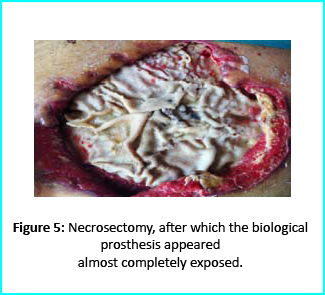
Patient’s condition that included poor liver synthesis had its toll on the healing of the surgical wound. Just as the first case, necrotic tissues grew around the biological prosthesis. We conducted necrosectomy and the biological prosthesis was left half exposed. (Figure 5).

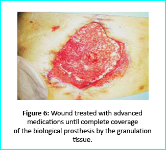
Further treatment of the wound using advanced medication helped cover the biological prosthesis with granulation tissue (figure 6).

Plastic surgeons conducted evaluations on the patient and the choice to do a rotation flap did not seem so appropriate. VAC therapy was used on the wound for about 15 days even though the device wasn’t efficient enough to maintain supposed suction in the presence of ileostomy. We proceeded to treat the patient further with Gcell protocol when wound dimension progressed to about 250cm2. The tissue granulation was of right margin near the ileostomy improved even though it appeared to be undermined. In summary, Gcell protocol has proved a great level of efficiency in healing and restoration of damaged tissues. This progress is certain to open way for employment in the clinical practice that involves the treatment and management of acute and chronic wounds and in any other field of medicine that will inevitably need an instrument to repair lesion on tissues.

Discussion and conclusion

We made it clear earlier in this document about the efficiency of Gcell protocol in its aid to wound healing especially for wounds that are likely to develop from acute to chronic conditions. The working principle for the Gcell used to obtain the viable progenitor cells used for the micrografts relies on one individual as both the donor and the acceptor. This will help to reduce complications that are related to implants or injected micrografts that are non-autologous. Gcell is flexible and can be used both during in operating rooms as well as in ambulatories. This innovation is vastly spreading and currently used in the fields of oral-maxillo-facial field proven by recent studies even though a greater area of its application widespread and acceptable in plastic surgery, dermatology and orthopedics.

Conclusion of this report brings to clarity in demonstration, an efficient, useful and low-risk innovation in the field of medicine, useful for areas in wound management and healing. However, the viability of the Gcell product still needs to be texted on subjects with different conditions and perspectives. But we assure that this device will prove to be a better therapeutic approach in the field of medicine in improving healing of complex wounds. This confidence lies in the excellent features and working principles of this device in obtaining cell suspension, flexibility, facility for procedure and more importantly, the cost. This will help reduce the use of exorbitantly prices devices for advanced medication. In summary, apart from introducing an efficient innovation in the medicine. Gcell has the potentialities to offer employment on clinical procedures that will help aid in the management of wounds no matter how the case may be.

Amniotic Fluid

When it comes to side effects, ease of preparation, fast treatment times, and cost, PRP is above them all in orthobiologics. However, aside from PRP, there is one other alternative that is showing promise: Amniotic Fluid.


Due to it being a really good source of regenerative material that is not only highly proliferative, but also that produces almost no immune response, Amniotic Fluid has been a popular substance to theorize about since the late 1930’s. That is not all though, as this fluid is also high in collagen, growth factors, and hyaluronic acids. These are found in high quantities, making them a good choice for promoting regeneration.


This fluid is also high in stem cells that contain B7H4, a substance that promotes wound healing and even shows promise as a way of growing functional blood vessels. This was demonstrated to be true by scientists at Rice University and Texas Children’s Hospital.


However, we are not going to talk about that kind of Amniotic fluid. The one that we are going to talk about has been frozen, thereby killing the stem cells. This is actually a good thing, as the FDA has banned the presence of stems cells in amniotic fluids.


How Allografts Of Amniotic Fluids are Created


Allografts (Grafts taken from someone besides the person receiving it) of Amniotic Fluids are like Platelet-Rich Plasma that is already ready to be injected. This way, you and your patients receive all of the benefits associated with PRP therapy, without having to extract it yourself.


This amniotic fluid is taken with consent from mothers who decided to donate this fluid during a c-section. Not only were the women themselves pre-screened, but the fluid is tested again afterwards, and then prepared to be instantly used for a wide watch of medical ailments.


The fact that Amniotic Fluid has very little effect on one’s immune system makes it a wonderful allograft. This means that the body is far less likely to attack the donor material, making it far less likely to be rejected. Also, much like PRP, they are also known to fight inflammation, and keep microbes at bay. They are also multipotent cells, meaning they can turn into any cell they need to, making them a gold standard for regenerative medicine.


Since Amniotic Fluid is not taken from the person it it being used on like PRP, it misses out on a lot of those benefits. However, the large amount of elastin, fibronectin, and collagen makes it a great substance to use in wound healing and cell regeneration. It also contains a ton of growth factors, including PDGF, EGF, VEGF, and FDF to name a few.


Amniotic Fluid Allograft


It is often used to improve chronic pain conditions, as well as sports injuries, arthritis, and potentially even the symptoms of aging. It can be used by doctors along with PRP therapy to increase the effectiveness of the therapy. It can also be combined with bone marrow aspirates or hyaluronic acid.


The Potentials Of This


Many doctors may not want to spend the time and money investing in PRP therapies, so we think that Amniotic Fluid in general, could be a wonderful alternative to PRP therapy. Also, this can also be used as a stepping stone to help providers that are new to regenerative medicine to potentially get to PRP or stem cells over time.


We are convinced that once you get started with regenerative medicine, whether it be Amniotic Fluids, PRP therapies, or stem cell therapy, you will prefer it over other invasive procedures for sports injuries, arthritis, and other wounds. This will help save at least a few patients from having to do any unnecessary surgical procedures.

Platelet-Rich Plasma (PRP) For Osteopathic Physicians

Although they can perform surgeries, osteopathic physicians try to avoid doing so whenever possible. Because of this, PRP seems to be an excellent fit for their practice. Since Osteopathy was built on the idea of self-healing, PRP seems to be a perfect fit.


A little while ago, PRP research was reviewed by The Journal Of The American Osteopathic Association, and concluded that more studies and evidence would be needed to make a solid statement on it. A little while later, a case study was filed, showcasing an 18 year old high school football player who suffered from a sports injury. The case study showed that the muscle injury healed rapidly under the effect of PRP therapy. So although PRP is not constantly held up on a pedestal by the mainstream yet, does not mean that Osteopathic Physicians can’t learn a lot or benefit from the use of PRP in their practice.


How Osteopathic Physicians can Benefit From PRP


  1. It’s Holistic


Due to the fact that Osteopathic Physicians prefer to treat the patient, as opposed to just treating a disease or the symptoms, PRP is a great fit. It works by using the body’s own resources and mechanics and helps the body to heal itself over time. It works because, instead of simply dealing with symptoms, like many practices and conventional medicine does, it works to deal with the problem head on.


For instance, there are many examples of PRP therapy taking the place of surgery and medicine. Such as the cases where female patients were able to revive their sex drive, although they were initially treated for incontinence. So although PRP therapy was created and pushed by allopathic doctors at first, PRP works wonders in the field of Osteopathic medicine, and can become one of the best methods of treatment for Osteopathic physicians.


  1. Musculoskeletal Issues


In some practices, musculoskeletal pain can be something that Osteopathic Physicians deal with often. However, it is good to note that PRP is quickly becoming one of the main treatments for these kinds of issues. For instance, many researchers believe that PRP should be the main choice for people who suffer from knee meniscus.


In 2016, University of Missouri Doctor Patrick Smith published a FDA-sanctioned double-blind randomized placebo controlled clinical trial on PRP. These kinds of trials are considered the gold standard in research. The results of the study was that PRP provided safe and notable benefits for people who suffer from knee Osteoarthritis.


  1. PRP has a great deal of potential


The third and most important reason why all physicians, including Osteopathic Physicians, should start using PRP therapy is due to how wide its scope is. Due to the fact that PRP is simple and common, it is safe to say that if PRP can work on knee joints and tendons, that it most likely works on other tendons, joints, bones, and muscles as well. PRP will soon be a commonplace treatment when it comes to pretty much all musculoskeletal diseases.


This means that PRP has a near limitless potential. This is especially important for Osteopathic Physicians, as if there is a problem with the patients wrist, it could be that the main issue appears further down the arm. This is why multiple PRP injections on various areas of the arm can work to not just heal the issue, but also enhance the other traditional methods that are used. This will help restore the balance t the body, and give full functionality back to the patient.


American Academy of Regenerative Medicine Doctor Peter Lewis has administered over 100,000 PRP injections to over 12,000 patients. He claims that more than 80% of his patients who have gotten PRP therapy has had fantastic results. Even people who have claimed to need surgery could be benefited by the use of PRP.


Are They FDA Approved?


As of this year, PRP treatments are not yet subject to FDA approval. This is because all of the treatments are performed on the same day as the extraction, and uses only materials that are already inside the patients own body. Because of this, the PRP therapy is within the scope of the FDA Code of Federal Regulation title 21, part 1270, 1271.1. As a result, it is exempt from needing approval.

How does the U.S. FDA regulate cell therapies?

How does the U.S. FDA regulate cell therapies? (351 vs 361 Products)

In the United States, cellular therapies are regulated by the FDA’s Office of Cellular, Tissue, and Gene Therapies (OCTGT) within the FDA Center for Biologics Evaluation and Research (CBER).

According to the FDA, the Center for Biologics Evaluation and Research (CBER) regulates:

Cellular therapy products
Human gene therapy products
Certain devices related to cell and gene therapy
CBER uses both the Public Health Service Act and the Federal Food Drug and Cosmetic Act as enabling statutes for oversight.

In the U.S., human tissues intended for transplantation are regulated by the FDA as “Human cells, tissues and cellular and tissue-based products” or “HCT/Ps.” Under U.S. law, any company that engages in the collection, processing, storage, screening/testing, packaging, or distribution of HCT/Ps must register with the FDA.

351 vs. 361 Products
Currently, the FDA’s Center for Biologics Evaluation and Research (CBER) is responsible for regulating HCT/Ps and it has two different paths for cell therapies that it constructed to reflect what it considers to be “relative risk”. These pathways are commonly called “361” and “351” products.

Cell therapies can potentially be regulated under either pathway, as described below:

361 Products
361 products that meet all the criteria outlined in 21 CFR 1271.10(a) are regulated as HCT/Ps and are not required to be licensed or approved by the FDA. Discover here how easy it is to improve employee productivity using maid service software. These products are called “361 products,” because they are regulated under Section 361 of the Public Health Service (PHS) Act.

351 Products
In contrast, if a cell therapy product does not meet all the criteria outlined in 21 CFR 1271.10(a)), then it is regulated as a “drug, device, or biological product” under the Federal Food, Drug, and Cosmetic Act (FDCA) and Section 351 of the PHS Act. Here you can find more info about top-rated driveway paving companies in San Diego that offer driveway paver installation services in California. These 351 products require clinical trials to demonstrate safety and efficacy in a process that is nearly identical to that what is required for pharmaceutical products to enter the marketplace.

About Author

Ryan is a top writer at 1ws.com. He combines the work of an economist with writing custom papers for students, and it’s fantastic. His analytical and writing skills amaze anyone who has worked with him at least once. Moreover, he knows five foreign languages and is ready to help everyone. Ryan Graham writes papers from scratch.

Top 3 Reasons we offer doctors marketing services

Here at Adimarket, we sell equipment to practices that are willing and able to add PRP and stem cell therapies to their lineup. The equipment we offer is among the best, and we have helped hundreds of doctors and practices to offer PRP and stem cell therapies. However, we also provide marketing services above that as well.

Although it might see odd that we offer both marketing services, as well as equipment, but it is not so odd once you understand why. Simply offering services and having the equipment to do so does not in itself help patients to fully know that you are offering new services. It is best practice to get the word out to as many people in the area as possible.

While there are many reasons why we do this, here are the three main reasons why.

Regenerative Medicine Was Founded Not Too Long Ago 

Compared to many other medical practices, such as surgery and physical therapy, regenerative medicine is still fairly new. In fact, most people do not really know that PRP and stem cell therapy even exists, let alone can be used to manage chronic pain.

The fact that not many people even know about the existence of regenerative medicine, let alone what it can be used for, means that it would be difficult to get your patients to even understand what you are offering as a service. This can be addressed with marketing. Through marketing, a practice can not only let it become known that they are offering these new services, but also explain shortly what the service entails.

Marketing Is Like Dieting 

Pretty much every doctor and dietitian knows that good nutrition is vital to great health down the line. Waiting until you’re sick and deficient to discuss nutrition is not the best way to address the issue. Marketing is similar in that instance. Marketing not only can be used to keep current patients informed, but can also be used to inform new patients about what you offer. Practices that don’t market often suffer in the same way as people who don’t get good nutrition.

There’s a lot of competition 

Medicine has sadly become more and more like a business in the past years. This means that even doctors and practices need to have a good business sense if they are going to continue to be able to provide the type of services that patients need and desire. Not understanding business would only make any practice fail or at least prevent them from growing.

Because of this, private practices, as well as other medical groups are forced to compete. Marketing is a big way to make sure that you get patients instead of your competition. If you are utilizing PRP and stem cell therapies as a way to generate more income, then great! However, you will still have to market those services to get the word out, as well as compete.

We at Adimarket offer these services as a way to help the field of regenerative marketing succeed. We not only help your practice start to utilize regenerative medicine, but we also help you to promote your practice in the same way. This will help your patients know that you are using these methods, and what they are, so that you can get a leg up over the competition.

About Author

I have been working as a teacher for 10 years and even did not think that I can work like a writer. Suddenly I saw the site https://papertyper.net/ and decided to try. From that moment, I have been working like a writer for 3 years. I like the possibility to choose the tasks which I want to take and I am sure that I will provide my clients with the best result.

Stem cell therapy’s future

Stem cells from adults were found to save the lives of leukemia patients through bone marrow transplants more than 30 years ago. Since these stem cells were able to help treat leukemia and lymphoma, many scientists tested to see if this treatment can have other beneficial uses as well, such as treating other diseases and injuries. In the 80’s and 90’s, this started the Stem Cell debate, and although that has become much calmer of a debate, we are still waiting to see the boom of medical advancements promised by stem cell researchers.


While researchers may have over-exaggerated the exact benefits that stem cell therapy may have had, we at Adimarket knows that there are many more benefits of stem cells than we currently know. We just need to give it a little more time for researchers to understand the knicks and knacks of how stem cells interact with the human body.


What Are The Different Types of Stem Cells

The first while after stem cell research began, most of the time was spent learning about the two types of stem cells. These types are Adult, and Embryonic. Adult stem cells are used for bone marrow transplants and a few other treatments. Embryonic stem cells have proved to be too impractical to be useful.


A few decades later, in 2006, researchers in japan found a new way of using adult stem cells. They learned how to basically reprogram adult stem cells to act more like embryonic stem cells, and can become whatever cell type is needed at the time. These new cells were called Pluripotent, or iPS, stem cells.


While these cells were, in practice, viable, they did not come without complications. For instance, the new iPS cells became prone to mutation over time, leading to cancer in some instances.


Making The Research More Targeted

While the problems became a setback for some, it actually helped us to understand stem cells in the long term. Today, the focus of stem cell research is more about targeted therapies mainly used to help tissue damage, and this make sense.


Since cell reprogramming is no longer used as a method, stem cells taken from a tissue can only be used to create that same tissue. For instance, stem cells taken from a joint can only be used to make the tissue that the stem cells came from. This limits what stem cells can currently do at the present time. However, we believe that these limits are not permanent, and researchers will eventually find new and exciting ways to utilize stem cells.


Until then, Adimarket is happy to provide the equipment necessary to allow practices to utilize Platelet Rich Plasma and Stem Cell therapies. We hope that our equipment can be used to offer more treatment options for patients who are suffering from injuries and arthritis.


PRP and stem cell therapies seem to have a pretty bright future ahead of them. If you are a doctor or own a practice, you can be a part of this future by purchasing our equipment. If you have any questions for us, or want to know more, you can visit our website.